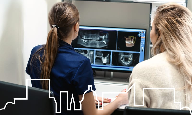
Staley Dental’s Guide to Understanding Dental X-Rays

Dental X-rays are an essential part of basic dental care.
In comic books, X-rays are touched by fantasy, allowing for all sorts of X-ray–based superpowers like Superman’s famous X-ray vision. While dental X-rays are less fantastical in real life, they’re still incredibly useful—even lifesaving. After all, sometimes we need to see what’s going on beneath the surface, and that’s where X-rays come in.
Since good portions of your teeth aren’t visible beneath your gum line, dental X-rays are also incredibly useful for your dentist. They’re so important that regular X-rays are considered a basic part of your regular dental care—they’re even covered by insurance. But what are X-rays? Are they really necessary? And what can they tell dentists about your oral health?
To help you understand these questions and more, we’ve put together a guide on everything you need to know about dental X-rays.
What are dental X-rays?
X-rays are a type of electromagnetic radiation—waves of energy that doctors and dentists use to create images of the inside of your body, particularly areas like your bones and teeth or the inside of your lungs. This works because X-rays are high energy with a short wavelength, allowing them to pass through objects much more easily than light does.
When X-rays are directed at you, they’re reflected by the parts of your body with low density, such as your muscles, fat, and skin, but absorbed by high-density parts, like bone and teeth. The result is a picture that shows your skin and muscles as dark, almost empty gray or black areas on the photo, while your bones and teeth show up a bright, contrasting white.
Modern X-rays can show a surprising amount of detail, from more obvious breaks to small hairline fractures along bones, or even pneumonia in your lungs, making it a powerful diagnostic tool.
How important are dental x-rays?
The incredible ability of X-rays to allow medical professionals to see what’s going on beneath the surface of your skin makes them a powerful diagnostic tool. A good portion of your teeth is located beneath the surface of your gums, so dental X-rays help dentists get a look at your tooth roots, their supporting structures, and your jaw to ensure that they’re healthy.
They’re able to spot issues, both above and below the surface of your gums, that may not be visible during a standard dental exam. As a result, dental X-rays can be used in a variety of ways, including diagnosing or examining issues like:
- Cavities, particularly those that are located between teeth or are too small for your dentist to notice with the naked eye.
- Dental abscesses or cysts.
- Damaged tooth roots.
- Impacted teeth.
- Tooth development in children.
- Bite alignment.
- Bone loss in your jaw.
- Failing dental restorations like a crown or filling.
- Tumors.
Beyond diagnosis, dental X-rays are also used to help plan dental treatments (like dental implants or bridges) and orthodontics (like braces or clear aligners). This makes X-rays beneficial in all stages of dental treatment, from prevention to diagnosis and even the treatment-planning process.
Why does my dentist take a couple of different kinds of X-rays?
There’s a surprising amount of variety when it comes to the types of dental X-rays you can get, but each one provides different benefits. There are two main categories of dental X-rays, intraoral and extraoral, both of which are taken digitally nowadays to provide a clearer image and instant results.
Intraoral X-rays
Intraoral X-rays are the most common type and are taken by placing an electronic sensor inside of your mouth. This sensor takes a digital image of your teeth and sends it directly to a computer screen so your dentist can look at it right away. Because of the electronic sensor’s placement in your mouth, intraoral X-rays focus on your teeth and jaws, showing them in great detail.
When you get a standard X-ray just to make sure your teeth and jaws are healthy, you’re likely getting an intraoral X-ray, such as a digital bitewing X-ray. Bitewing X-rays are taken regularly to ensure that your teeth are healthy because they provide a detailed look at your entire tooth from the crown to the roots, and even the bone supporting your teeth. They’re incredibly good at detecting issues like decay between your teeth, failing fillings, or bone loss, as well as for planning dental restorations like implants, bridges, or crowns.
Extraoral X-rays
Extraoral X-rays are the second category of X-rays, where the sensor is located outside of your mouth. These X-rays do show teeth, but not in as much detail as intraoral X-rays, as their main focus is capturing images of your jaws and skull. Panoramic X-rays are a common type of extraoral X-ray that’s taken as a single, continuous picture using a machine that rotates around your head.
This type of X-ray is generally only taken when your dentist decides you need it, but they’re incredibly useful for keeping an eye on the development of wisdom teeth, identifying impacted teeth or cysts, diagnosing issues with your TMJ or sinuses, planning restorative dentistry treatments, and more.
Why does my dentist sometimes have me bite down on bitewings?
The little plastic tabs (bitewings) that you bite down on during an intraoral X-ray have a two-fold purpose. They’re the sensor that takes the place of traditional film and helps take the picture, giving a clear image of your teeth and jaw. They also help the technician line up the machine and keep your teeth still and steady while the X-ray is being taken. This allows for a better, more detailed photo in fewer tries, saving you time and getting your results to you sooner.
How safe are dental X-rays?
Although X-rays do produce radiation, they produce a very small amount of it. The modern digital X-rays we use produce even less radiation than traditional film X-rays, in part because the image is taken much more quickly. You’ll also wear a lead apron shield during your X-ray to limit your exposure even further. As a result, digital X-rays expose you to only a very tiny amount of radiation.
We’re all exposed to natural levels of radiation from the environment around us every day, and the amount of radiation from your X-ray is about equivalent to the natural radiation you encounter on an average day. It’s completely safe—more than that, the benefits provided by the X-ray’s diagnostic capabilities more than outweigh any perceived risks.
How often are dental X-rays needed?
Standard bitewing X-rays are generally taken once or twice a year to check up on the health of your teeth and gums. This gives Dr. Staley the best chance of spotting issues early that can’t be seen during a standard exam. Exactly how often you need these X-rays depends on your oral health history and risk factors. For example, if you have a history of oral health issues or have risk factors that make you more vulnerable to cavities or gum disease, you might need a bitewing X-ray once a year just to make sure your oral health stays in great shape.
Panoramic X-rays, however, are needed much less often, and usually only when a specific issue needs to be diagnosed or checked up on. You may also get a full mouth series of dental X-rays if you’re a new patient, as it helps our team assess your overall oral health and provides an initial set of images to refer to while we treat you. A full series like this lasts about three to five years before it needs to be redone.
Taking a dental X-ray is a quick, easy, and safe process. The detailed images they create allow dentists to detect oral health issues early and make it easier to plan treatments, making them an essential part of keeping your teeth, gums, and jaws healthy in the long term. If you’d like to learn more about dental X-rays or you’re due for a checkup, feel free to schedule a consultation with Dr. Staley at any time.